What is MRI ?
MRI stands for Magnetic Resonance Imaging. It is a non-invasive radiology imaging technology that uses magnetism, radio waves, and a computer to produce detailed anatomical images. It is often used for disease detection, diagnosis, and treatment monitoring. Some applications may include MR Angiography, Functional Imaging and Perfusion / Diffusion scanning.
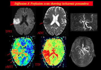
Perfusion/ Diffusion scanning
|
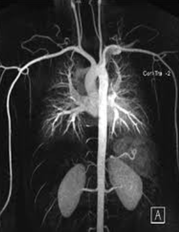
MR Angiography
|
Before the MRI technology, x-rays and CT scans were commonly used to image the body. In the case of x-rays, the overall contrast resolution of an x-ray image is poor and the image formed is gray and flat. With CT scanners an image can be produced with more contrast than x-rays, which helps in detecting malignant tissue. The principle advantage of MRI over x-rays and CT scan is its excellent contrast resolution. Generally, x-rays and CT scans are used to visualize bone structures. Now, MRI is widely used as it is able to effectively detect minute contrast differences in soft tissue and detect soft tissue lesions. Additionally, it is able to create images in all planes.
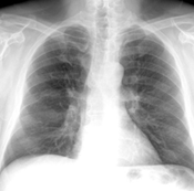
X- Ray: of the rib cage
|
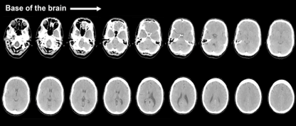
CT Scan: of the brain
|
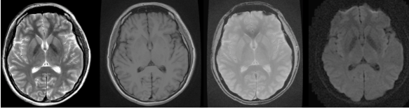
MRI : of the brain
